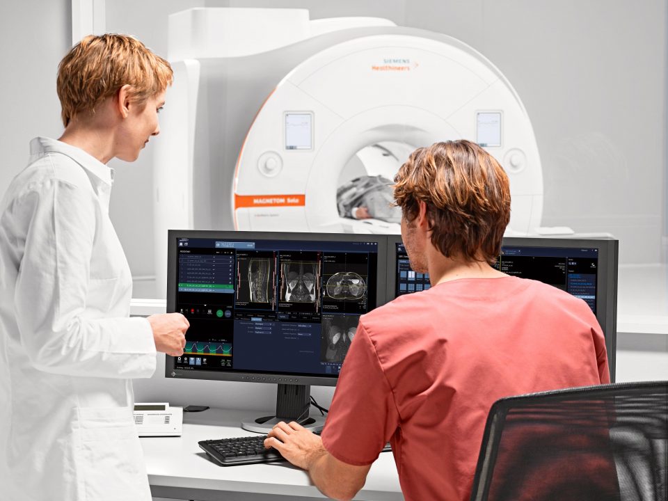
Full-Service Imaging
Conveniently Located on our Browns Mills Campus
- Same Day and Next Day Appointments
- No Long Waits
- All Major Insurances Accepted
Pediatric Echocardiography
A pediatric echocardiogram is a test performed on children that uses sound waves to create pictures of the heart. It is used to help diagnose defects of the heart that are present at birth.
This test is done to examine the function, heart valves, major blood vessels, and chambers of a child’s heart from outside of the body.
How does it work?
During the procedure:
- The sonographer puts gel on the child’s ribs near the breastbone in the area around the heart. A hand-held instrument, called a transducer, is pressed on the gel on the child’s chest and directed toward the heart. This device releases high-frequency sound waves.
- The transducer picks up the echo of sound waves coming back from the heart and blood vessels.
- The echocardiography machine converts these impulses into moving pictures of the heart. Still pictures are also taken.
- Pictures can be two-dimensional or three-dimensional.
- The entire procedure lasts for about 20 to 40 minutes.